
Files
Download Full Text (1.0 MB)
Publication Date
4-29-2020
Disciplines
Internal Medicine
Description
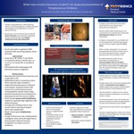
When Less Virulent Becomes Virulent!!
An Atypical Presentation Of Streptococcus Viridians
Samreen Kahn, MBBS
Providence Portland Medical Center – Portland, OR
Additional Authors: Stavan Patel MS, MD
Introduction: There are several cardiac and non-cardiac causes and risk factors for the development of infective endocarditis (IE) in young healthy adults. Some risk factors include prior IE, history of valvular or congenital heart disease, IV drug use, indwelling intravenous lines, immunosuppression, or a recent dental or surgical procedure. Most two common sites of IE are mitral and aortic valve. We present a case of a 45-year-old man with no-known risk factors, who presented with respiratory distress and was found to have streptococcal viridians (SV) endocarditis.
Case Presentation: A healthy 45-year-old male presented with a four-day history of acute dyspnea and new-onset of fevers and chills for 24 hours before arrival. He denied any chest pain, dizziness, palpitations, LOC, weakness, increasing LE edema, or palpations. Patient denied any recent travels, prolonged non-ambulatory state, recent sick contacts, IV drug abuse, high risk sexual activities, no-known personal cardiac history, no recent dental manipulation. While in ED, he reported new onset of chest heaviness, and a non-productive cough. Vitals: T 37.2 C, BP 125/9, P 122, RR 26. EKG showed sinus tachycardia with non- specific ST&T wave abnormalities/No prior ECG where available to compare. Troponin 64 ng/L. ABG pH 7.230, PCO2 27.4 mmHg, PO2 82.1 mmHg. A-a gradient calculated to be greater than 200. CXR showed pulmonary vascular congestion. CTPA for PE was deferred as patient could not remain supine during examination secondary to worsening respiratory status. Patient was moved to medical ICU, with rapid worsening of respiratory status requiring intubation. Urgent bronchoscopy revealed diffuse alveolar hemorrhage consistent with severe acute respiratory distress syndrome (ARDS) with multifocal pneumonia. On initial presentation IE was not excepted and patient was treated for viral and bacterial causes of ARDS. Echocardiogram showed aortic valve insufficiency with questionable vegetation with leaflet disruption. Subsequently blood cultures showed gram-positive cocci. Given the combination of aortic insufficiency (AI) and gram-positive cocci IE speculated which was soon followed by aortic valve replacement with intraoperative finding of aortic intra-annular abscess. Clinical course was complicated with cardiogenic shock and multi-organ failure. Blood cultures subsequently grew SV.
Case Discussion: Despite advances in medical, surgical and critical care interventions, IE remains a life-threatening illness. SV is not uncommon but are routinely seen in those with underlying heart disease and dental manipulation. SV is responsible for 40-60% of IE in normal valves [3, 4] and patients (young males and over 45 years of age) usually with mitral valves. It is commonly associated with heart failure and lesion such as peri-annular abscesses, fistulas, or pseudo-aneurysms with risk of mortality at 15% [1, 2]. Diagnosis may be difficult given no risk factors, non-specific symptoms but does not exclude this pathogen as a cause.
Department
Internal Medicine
Department
Graduate Medical Education
Conference / Event Name
Academic Achievement Day, 2020
Location
Providence Portland Medical Center, Internal Medicine Residency, Portland, Oregon
Recommended Citation
Khan, Samreen; Patel, Stavan; and Nidiry, Mary Anne, "When less-virulent becomes virulent!! An atypical presentation of Streptococcus Viridians." (2020). Providence Portland Medical Center Internal Medicine 2020. 16.
https://digitalcommons.providence.org/ppmc_internal/16


