
Files
Download Full Text (1.3 MB)
Publication Date
5-2021
Keywords
oregon; portland; ppmc
Disciplines
Internal Medicine
Abstract
Introduction Aspergillus spores are routinely inhaled.Innate immune defenses prevent fungal growth and disease in immunocompetent individuals.However, immunocompromised patients are at risk of developing invasive aspergillosis. Diagnosis of invasive aspergillus is often difficult as biopsy is not always feasible and relies on the interpretation of non-invasive testing. Early recognition and initiation of therapy is paramount as the mortality rate is high.
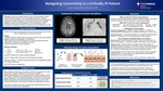
Case Presentation Patient is a 59 year oldfemale with a history of alcohol abuse admitted with acute encephalopathy and new onset seizure. History: -Initially presented 3 weeks earlier for jaundice, found to have acute alcoholic hepatitis, started and discharged on prednisolone -Now presenting with new onset grand mal seizure and acute confusion -Obtunded on admission and admitted to the ICU -Required intubation for airway protection Admission Workup: Chest XR: Diffuse ground glass opacities bilaterally Head CT: Patchy white matter foci in the left frontal and parietal MRI Brain (Image 1): T2/FLAIR hyperintense lesions in frontal/parietal regions in a scattered distribution TTE: EF 75% and no vegetations ALT 88 AST 140 Bilirubin 19.7
Hospital Course Day 1: Started on Vancomycin and Zosyn. TEE with no vegetations. Unable to perform LP. Unable to liberate from mechanical ventilation Day 4: Repeat MRI brain with increased size and number of lesions. Chest CT (Image 2): Multifocalair disease bilaterally with ill-defined left lower lobe density. Respiratory cultures growing aspergillus. Started on IV voriconazole Day 8: Galactomannan and beta-D-glucan positive. Repeat head CT with increased CNS lesions. Transitioned to comfort care after family discussion. Autopsy with multiple CNS fungal abscesses with leptomeningeal extension, consistent with disseminated aspergillosis
Discussion When to Test for Invasive Fungal Infections Testing for invasive fungal infection should be initiated promptly in all patients who have risk factorsand the differential includes fungal etiology. Radiographic Characteristics Pulmonary Aspergillosis:1. Single or multiple nodules (+/-cavitation) 2. Patchy/segmental consolidations 3. Peribronchial infiltrates (+/-tree-in-bud) CNS Aspergillosis:1. Ring-enhancing lesions 2. Cerebral cortical/subcortical infarction +/-hematoma 3. Sinus involvement with CNS extension How to Interpret Fungal Biomarkers and Cultures Beta-D-Glucan:Cell wall component in fungi. Found on all fugus and not specific for aspergillus. (+) test may indicate fungal infection, but not specific Galactomannan:Polysaccharide on aspergillus walls. More specific for aspergillus and can be diagnostic in the correct context. Sensitivity decreased with antifungal therapy and false (+) can occur with Zosyn. If serum results are (-), BAL can be performed, which provides additional sensitivity. Fungal Cultures:Does not always mean active infection. We are constantly inhaling conidia. (+) cultures need to be weighed again the probability of aspergillosis as the cause of disease. Biopsy is the gold standard, but often cannot be obtained in critical illness
Conclusions Invasive aspergillosis often presents with non-specific symptoms and knowing the risk factors and radiographic findings can help identify who to test -Diagnosis is based upon both identifying the organism via biopsy or biomarkers as well as probability that aspergillus is the cause of disease -Positive sputum fungal stain/culture or positive galactomannan should prompt therapy in the setting of high clinical suspicion and risk factors for aspergil
Specialty/Research Institute
Internal Medicine
Specialty/Research Institute
Graduate Medical Education
Conference / Event Name
Academic Achievement Day, 2021
Location
Providence Portland Medical Center


